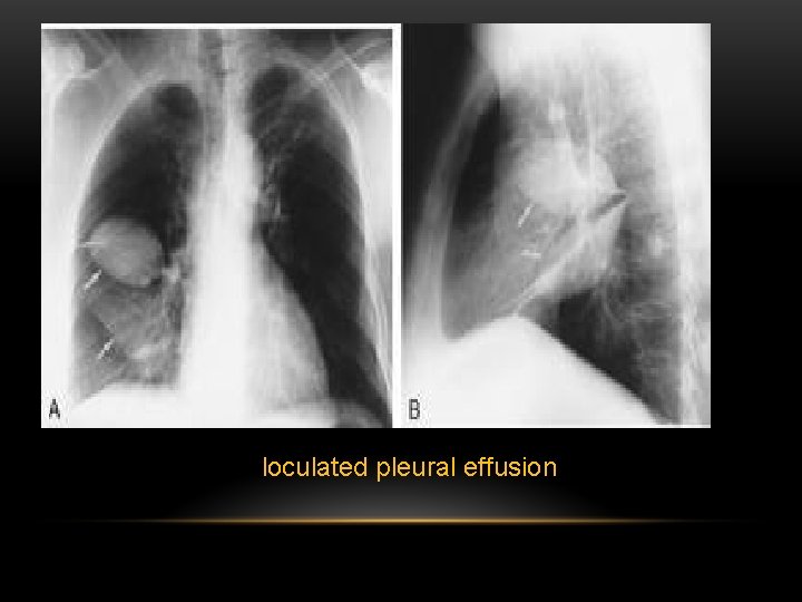
Loculated Pleural Effusion : Loculated Pleural Effusion Radiology Case Radiopaedia Org. Conventional chest radiography and computed tomography (ct) scanning are the primary imaging modalities that are used for evaluation of all types of pleural disease, but ultrasound and magnetic resonance. Pleural effusion occurs when fluid enters the lining of the lungs. Surgically implanted pleuroperitoneal shunts are another treatment option for recurrent, symptomatic effusions, most often in the setting of malignancy, but. Loculated pleural effusion masquerading as mediastinal tumour had been reported but pleural effusion that conformed to the contour of a lung lobe is rare. The anatomy (a) of the pleural effusion is based on the size of the effusion, whether it is free flowing, and whether the parietal pleural is thickened.
Complex septated, complex nonseptated, or homogeneously echogenic effusions are always exudates (fig. Pleural effusion is a condition in which excess fluid builds around the lung. Pleural effusions in the intensive care setting. If you are struggling with chest pain that gets worse when you cough or inhale, chances. This case highlights the atypical but unique presentation of a transudative pleural effusion and demonstrates the risk of repeated thoracocentesis complicating a simple clinical presentation.
Pdf Intrapleural Streptokinase For Tuberculosis Loculated Pleural Effusion Semantic Scholar from d3i71xaburhd42.cloudfront.net If you are struggling with chest pain that gets worse when you cough or inhale, chances. Of loculated pleural effusions* jeffreys. The etiology of the pleural effusion determines other signs and symptoms. Pleural effusions in the intensive care setting. Encysted pleural fluid is visualized between the right upper and middle lobe (s). Empyema is defined by purulent fluid collection in the pleural space, which is most commonly caused by pneumonia. Conventional chest radiography and computed tomography (ct) scanning are the primary imaging modalities that are used for evaluation of all types of pleural disease, but ultrasound and magnetic resonance. Most malignant effusions can be controlled by thoracentesis and/or closed thoracostomy tube drainage and sclerosis of the pleural cavity.
In general, pleural effusions can be divided into transudates (caused by fluid leaking from blood vessels) and exudates (where fluid leaks from inflammation of the pleura and lung).
Icu patients cannot sit up and the effusion layers posteriorly. Most effusions start like this and can be easily missed. Treatment may fail if the catheter is not placed optimally within the loculation or if the fluid is hemorrhagic or fibrinous. Most malignant effusions can be controlled by thoracentesis and/or closed thoracostomy tube drainage and sclerosis of the pleural cavity. The largest pocket of fluid is present posteriorly at the right lung base, with associated atelectasis and minor consolidation. The pleural fluid is called a transudate if it permeates (transudes) into the pleural cavity through the walls of intact pulmonary vessels. What are the different appearances of pleural effusion? Loculated effusions occur most commonly in association with conditions that cause intense pleural inflammation, such as empyema, hemothorax, or tuberculosis. The etiology of the pleural effusion determines other signs and symptoms. Loculated malignant effusions however, are inherently resistant to the usual approaches because of nonexpanding underlying lung. Pleural effusion is an accumulation of fluid in the pleural space that is classified as transudate or exudate according to its composition and underlying pathophysiology. Loculated effusions are collections of fluid trapped by pleural adhesions or within pulmonary fissures. Pleural effusion occurs when fluid enters the lining of the lungs.
What Is Loculated Pleural Effusion from slidetodoc.com This case highlights the atypical but unique presentation of a transudative pleural effusion and demonstrates the risk of repeated thoracocentesis complicating a simple clinical presentation. Diffuse nodules and opacification in right lung with compressive atelectasis. Icu patients cannot sit up and the effusion layers posteriorly. An anechoic effusion can be a transudate or exudate (fig. The pleural fluid is called a transudate if it permeates (transudes) into the pleural cavity through the walls of intact pulmonary vessels. If you are struggling with chest pain that gets worse when you cough or inhale, chances. Complex septated, complex nonseptated, or homogeneously echogenic effusions are always exudates (fig. Empyema is defined by purulent fluid collection in the pleural space, which is most commonly caused by pneumonia.
Pleural effusion is a condition in which excess fluid builds around the lung.
If you are struggling with chest pain that gets worse when you cough or inhale, chances. What are the different appearances of pleural effusion? Left pleural effusion with high density material at the posterior costophrenic angle. Pleural effusion affects more than 1.5 million people in the united states each year and often complicates the management of heart failure, pneumonia, and malignancy. Of loculated pleural effusions* jeffreys. Most malignant effusions can be controlled by thoracentesis and/or closed thoracostomy tube drainage and sclerosis of the pleural cavity. The anatomy (a) of the pleural effusion is based on the size of the effusion, whether it is free flowing, and whether the parietal pleural is thickened. Pleural fluid is seen extending to the right oblique fissure. Pleural effusions in the intensive care setting. When a pleural effusion is loculated, the standard treatment methods of intercostal tube drainage and pleurodesis may not be helpful. Loculated malignant effusions however, are inherently resistant to the usual approaches because of nonexpanding underlying lung. Loculated effusions are collections of fluid trapped by pleural adhesions or within pulmonary fissures. Icu patients cannot sit up and the effusion layers posteriorly.
Empyema is defined by purulent fluid collection in the pleural space, which is most commonly caused by pneumonia. A pleural effusion is due to the manifestations of another illness.; Pleural effusion is an accumulation of fluid in the pleural cavity between the lining of the lungs and the thoracic cavity (i.e., the visceral and parietal pleurae). Treatment may fail if the catheter is not placed optimally within the loculation or if the fluid is hemorrhagic or fibrinous. It was successful in breaking the locules and draining the effusion.
In general, pleural effusions can be divided into transudates (caused by fluid leaking from blood vessels) and exudates (where fluid leaks from inflammation of the pleura and lung).


Post a Comment for "Loculated Pleural Effusion : Loculated Pleural Effusion Radiology Case Radiopaedia Org"