Tendon Diagram / 2 808 Tendon Vector Images Free Royalty Free Tendon Vectors Depositphotos. Attaches the calf muscles to the calcaneus, most important muscles for running, jumping, walking etc. Diagram of tendons in forearm. It attaches to the wrist bone, the pisiform, and as well as the 5th hand bone. Foot anatomy diagram, foot joint diagram, foot sprain diagram, foot tendons and ligaments pain, leg tendon diagram. A tendon is a band of tissue that connects a muscle to a bone.
If you tear the biceps tendon at the shoulder, you may lose some strength in your arm and have pain when you forcefully turn your arm from palm down to palm up. Fpe medical review board a foot pain diagram is a great tool to help you work out what is causing your ankle and foot pain. Attaches the calf muscles to the calcaneus, most important muscles for running, jumping, walking etc. Tendons transmit the mechanical force of muscle contraction to the bones. Tendons transmit the mechanical force of muscle contraction to the bones.
Tendons In The Foot Tendonitis Remember To Stretch Feet Too Properly Before Exercising Especially If You Are To Wal Tendonitis Foot Ankle Anatomy Anatomy from i.pinimg.com Tendons, located at each end of a muscle, attach muscle to bone. Tendon diagrams and design force vectors. The two peroneal tendons in the foot run side by side behind the outer ankle bone. The pubis, ischium, and ilium together constitute the pelvis while the thigh bone is the femur. The ecu tendon works along with the ecrl and ecrb to straighten the wrist. The changes in ligaments and tendons generally occur more slowly than adaptation in bone, because ligaments and tendons have less vascular supply. When the muscles tighten (contract) arguably, the most important tendon is the achilles tendon, which allows the calf muscles to move. Your biceps tendons attach the biceps muscle to bones in the shoulder and in the elbow.
A tendon is a tough yet flexible band of fibrous tissue.
Anatomy of leg muscles and tendons anatomy diagram leg muscles and tendons anatomy diagram pics photo, anatomy of leg muscles and tendons anatomy diagram leg muscles. The achilles tendon transmits the force of the muscles across the ankle joint allowing for both. Its muscle belly is in the forearm. Tendon diagram simple / 8.4c: Tendons transmit the mechanical force of muscle contraction to the bones. Bones, cartilage, ligaments, and tendons. This tendon connects the patella (kneecap) to the tibia. Tendon diagrams and design force vectors. In the back and elsewhere in the body, tendons attach muscles to bones. Diagram of inside the body. Again, our knowledge of how mechanical stimulus mediates ligament and tendon structure is more empirical and less. Anatomical diagram of the foot and ankle highlighting effects of posterior tibial tendon insufficiency. The fcu tendon is one of two tendons that bend the wrist.
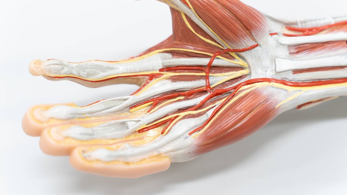
If the tendon cannot be identified then a complete tear of the tendon should be sought. Tendons are the connection between bones and muscles. Tendon diagrams and design force vectors. Tendons are the connection between bones and muscles tendon diagram. Diagram of tendons in forearm.
Tendon Anatomy Physiopedia from swiftype-ss.imgix.net When the muscles tighten (contract) arguably, the most important tendon is the achilles tendon, which allows the calf muscles to move. Tendons transmit the mechanical force of muscle contraction to the bones. If you feel the outside of your knee you'll feel this tendon. The knee joint is a complex structure that involves bones. Foot anatomy diagram, foot joint diagram, foot sprain diagram, foot tendons and ligaments pain, leg tendon diagram, peroneal tendonitis, foot, foot anatomy diagram, foot joint diagram, foot sprain diagram, foot tendons and ligaments pain, leg tendon diagram, peroneal tendonitis. The achilles tendon is also called the calcaneal tendon. The bones of the hip include the femur, the ilium, the ischium, and the pubis. They are attached to the femur (thighbone), tibia (shinbone), and fibula (calf bone) by fibrous tissues called ligaments.
Diagram of tendons in forearm.
Also allows the action of raising up onto toes. Diagram of inside the body. In the back and elsewhere in the body, tendons attach muscles to bones. The achilles tendon is the largest. Attaches the calf muscles to the calcaneus, most important muscles for running, jumping, walking etc. On the other hand, the insertion is where a tendon attaches that muscle to the *more* movable bone. Learn about the anatomy and physiology of tendons. Below is a diagram of the hamstring tendon. The achilles tendon is the strongest and largest tendon in the body. Numerous muscles help stabilize the three joints of. Your biceps tendons attach the biceps muscle to bones in the shoulder and in the elbow. This hd wallpaper knee diagram tendons has viewed by 709 users. It attaches to the wrist bone, the pisiform, and as well as the 5th hand bone.
If the tendon cannot be identified then a complete tear of the tendon should be sought. Tendons are the connection between bones and muscles. Allows the action of raising the foot. The rotator cuff is a group of four muscles and tendons that surround the glenohumeral joint. Numerous muscles help stabilize the three joints of.
Torn Horse Tendon The Long Road Back From This Equine Injury Expert How To For English Riders from practicalhorsemanmag.com You can see a diagram of the achilles tendon below. Also allows the action of raising up onto toes. The tendon travels along the inside of the forearm on the side of the small finger and crosses the wrist. On the other hand, the insertion is where a tendon attaches that muscle to the *more* movable bone. When the muscles tighten (contract) arguably, the most important tendon is the achilles tendon, which allows the calf muscles to move. This results in collapse of the arch of the foot (commonly called flatfoot or flat foot), along with foot and sometimes ankle deformities that can become debilitating or disabling in later stages. Diagram of inside the body. They are attached to the femur (thighbone), tibia (shinbone), and fibula (calf bone) by fibrous tissues called ligaments.
The achilles tendon is a tough band of fibrous tissue that connects the calf muscles to the heel bone (calcaneus).
The knee joint is a complex structure that involves bones. The achilles tendon is also called the calcaneal tendon. Fpe medical review board a foot pain diagram is a great tool to help you work out what is causing your ankle and foot pain. Tendons are found throughout the body, from the head and neck all the way down to the feet. The fcu tendon is one of two tendons that bend the wrist. The achilles tendon enables us to walk, without it we would not be able to raise our heels of the ground. A tendon is a band of tissue that connects a muscle to a bone. Black and white print showing the musculoskeletal system of a human hand, including the bones, muscles, cartilage, tendons, ligaments, and joints,. Ligaments and tendons are adapted in response to changes in mechanical stiffness. A muscle's origin is where a tendon attaches it to the *less* movable bone. When the muscles tighten (contract) arguably, the most important tendon is the achilles tendon, which allows the calf muscles to move. The tendon connects muscle to the bone. Below is a diagram of the hamstring tendon.
Share :
Post a Comment
for "Tendon Diagram / 2 808 Tendon Vector Images Free Royalty Free Tendon Vectors Depositphotos"


Post a Comment for "Tendon Diagram / 2 808 Tendon Vector Images Free Royalty Free Tendon Vectors Depositphotos"